CANTER - Center for Applied Tissue Engineering and Regenerative Medicine
Laboratory Homepage

Description
Rooms: C200, C 202, D 401, D104 – D106b
Phone: -3671

The following devices are currently available on the CANTER:
- Basic cell biology equipment: safety workbenches for cell culture (S1, S2), incubators, autoclaves, cell microscopes, etc.
- Basic molecular biology equipment: centrifuges, multiplate readers, gel documentation
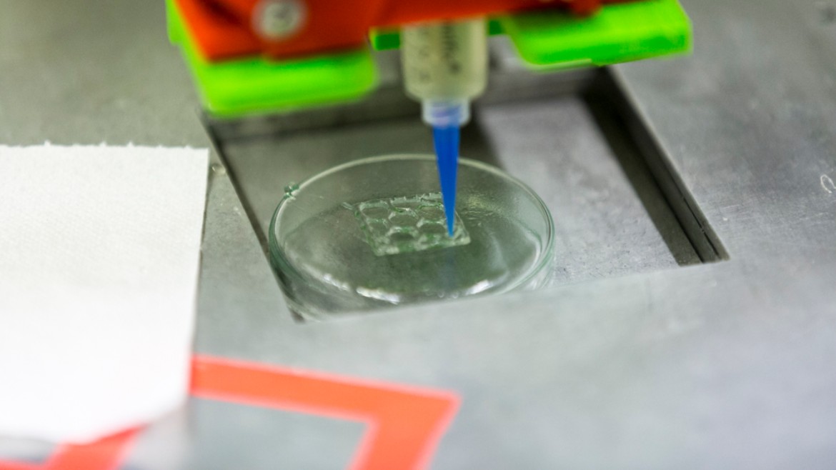
- 3D printer (Ultimaker 2, Form 2)
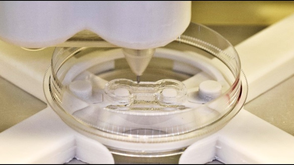
- Multiple bioprinters for printing cell-laden hydrogels (in-house development)
- Cryotoma for the preparation of tissue samples
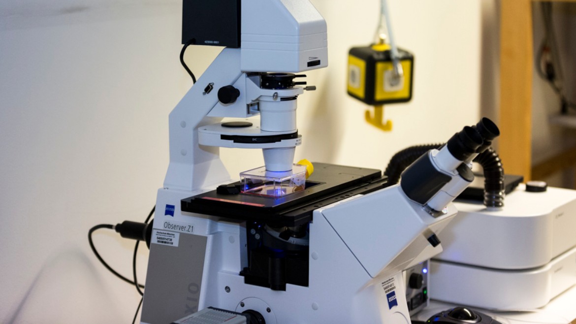
- Inverse fluorescence microscope with structured illumination for live cell imaging (time laps)
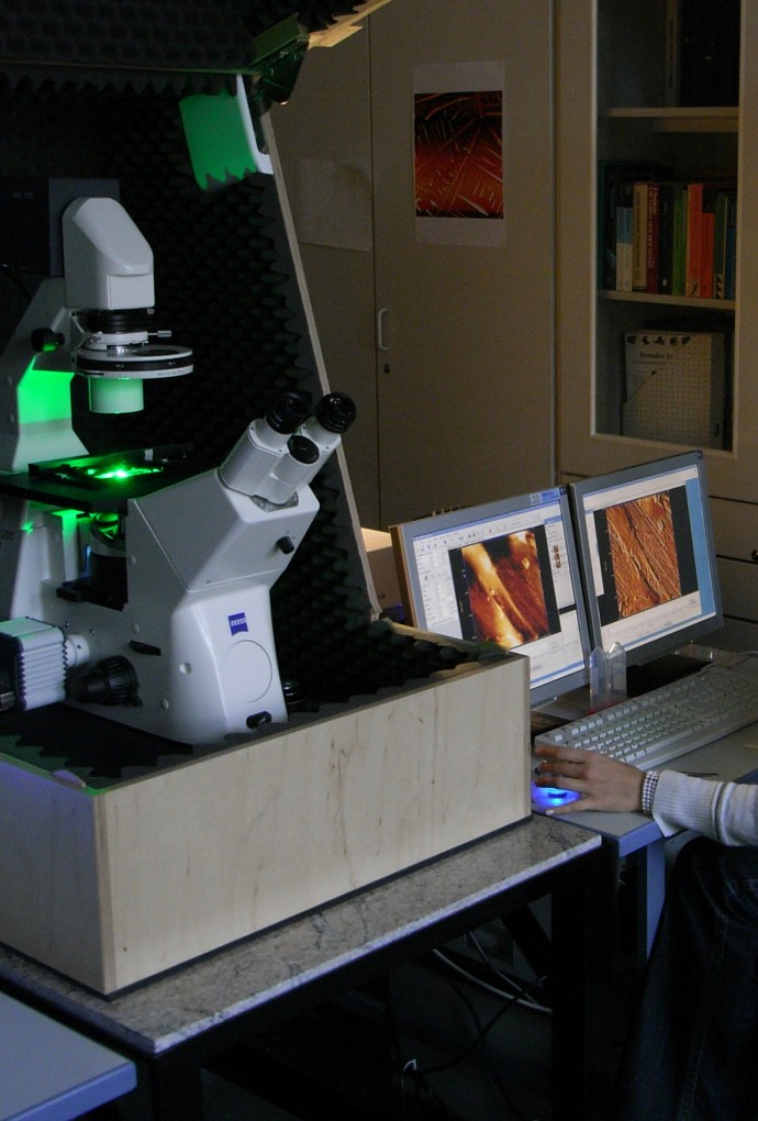
- Scanning force microscopes (Nanowizard, Nanoscope IIIa, MFP 3D), partly coupled with inverse fluorescence microscopes
- Micro-Univers Tester (Mach 1)
- In addition, other methods and devices are available in the laboratories involved in THE CANTER: a wide range of molecular and cell biology techniques, other 3D printers, including a 2-photon stereolithography printer, and a Laser sintering system, traught testing machines, confocal, SHG and STED microscopes, a scanning electron microscope, and powerful calculators for simulation
In addition, other methods and devices are available in the laboratories involved in THE CANTER: a wide range of molecular and cell biology techniques, other 3D printers, including a 2-photon stereolithography printer, and a Laser sintering system, tensile test equipment, confocal-, SHG- and STED- microscopes, a scanning electron microscope, and powerful calculators for simulation

The aim of the CANTER is to develop technologies based on the understanding of structural-function relationships in healthy and pathological tissues that make it possible to produce tissue replacement materials in the laboratory that are individually tailored to the needs of the patients. In addition to cell and molecular biology methods, generative manufacturing processes and materials technology, analytical methods are also required to examine tissues and tissue constructs with regard to their microscopic structure and biological functionality. The CANTER therefore conducts project, final and doctoral theses of students and graduates of the Bachelor's and Master's programmes in Bioengineering, Mechatronics, Physical / Chemical Engineering, Micro- and Nanotechnology, Photonics and Human Medicine. The topics range from the production and testing of bioinks, to the development of new 3D printing processes and bioreactors, to the biophysical characterization of cells and tissues.
Currently, work is being filled in the following areas:
- Optimization of hydrogels for 3D printing or shedding of cell-laden gels as well as molecular and cell biological characterization of the printed cells
- Development of bioreactors for 3D cell culture
- Characterization of the structure and mechanical properties of healthy and pathological tissues by means of scanning force microscopy
Cooperative promotions:
- Production of a 3D tissue model for the study and targeted stimulation of cell migration and cell growth along E-module gradients of the extracellular matrix, Amelie Erben, TU Munich, current
- Development of a 3D-printed microfluidic for the analysis of the influence of nanoscale slats on fluid dynamics in blood vessels, Benedikt Kaufmann, TU München, current
- Investigating the effect of alpha10 integrin and heperan-sulfates on the biomechanical properties and regeneration potential of articular cartilage using atomic force microscopy, Lutz Fleischhauer, LMU, current
- Determination of the modulus of elasticity on intact and degenerated cartilage by nano- and microindentation measurement, Bastian Hartmann, LMU, current
- Laser-induced transfer of human mesenchymal cells using near infrared femtosecond laser pulses for the precise configuration of cell nichoids, Jun Zhang, Universität Regensburg, current
- Integration of laser doppler vibrometry into the bioprinting process for the generation of three-dimensional bioactive constructs, Sascha Schwarz, TU München, current
- Mechanotransduction on the single cell level using AFM based single cell force spectroscopy (SCFS) for mechanical stimulation, Stefanie Kiderlen, LMU-München, current
- Cultivation systems for 3D cultures in tissue engineering, Jakob Schmid, LMU Munich, current
- Streptococcus pneumoniae TIGR4 pilus-1 biogenesis and functional-biomechanical aspects of adhesion molecule RrgA during interaction with the host, Tanja Becke, TU-München, current
- Investigation of the molecular and biophysical mechanisms of prostate cancer cell interactions with bone marrow-derived proteins and stem cells, Ediz Sariisik, LMU-München, concluded 2016
- Structure-function relationship of the cartilage extracellular matrix assessed by atomic force microscopy, Carina Prein, Ludwig-Maximilians-Universität München, concluded 2016
- Mechanochemical Stability of Covalent Bonds – Investigation with Atomic Force Microscope and Quantum Chemical Calculations, Michael Pill, Christian-Albrechts-Universität at Kiel concluded 2015
- Single-Molecule Force Spectroscopy of Covalent Bonds – Comparison of the Dynamic and the Force-Clamp Approach, Sebastian Schmidt, Christian-Albrechts-Universität zu Kiel, concluded 2011

The CANTER was founded in 2011 as a cooperation project of LMU, HM and TUM. It is based on an established cooperation network, of engineers, physicians and natural scientists with joint research projects, courses and jointly supervised thesis and doctorates. The participating scientists include:
- A. Aszodi, ExperiMed, LMU
- W. Böcker, General, Accident and Recovery Surgery, LMU
- H. Clausen-Schaumann, Biophysics and Nanoanalytics, HM
- D. Docheva, Laboratory for Experimental Accident Surgery, University of Regensburg
- T. Hellerer, Multi-Photon-Imaging Lab, Biophotonics, HM
- H. Huber, Lasercenter, HM
- R. Huber, Bioprocess Engineering, HM
- K. Pham, Materials Technology Lab, HM
- HG. Machens, Klinik und Poliklinik für Plastische Chirurgie und Handchirurgie, KRI, TUM
- J. Roths, Photonics Lab, HM
- M. Schieker, ExperiMed, LMU
- A.F.Schilling, Department of Accident Surgery, Orthopaedics and Plastic Surgery, UMG
- E. Steinhauser, Materials Technology Lab, HM
- S. Sudhop, CANTER, HM
- C. Tille, Composite Laboratory Design and CAx (KCA), HM
CANTER cooperates with companies in the fields of biotechnology/nanobiotechnology, materials technology/biomaterials, microsystem technology, sensor technology, optics/laser technology, engineering and medical technology.
Scientists of the CANTER are involved in numerous research projects, among others in the research heavyweight production and biophysical characterization of three-dimensional tissues funded by the Bavarian Ministry of Science and the DFG Research unit FOR 2407 Exploring articular cartilage and subchondral bone degeneration and regeneration in osteoarthritis - ExCarBon.
J. Roths, H. Clausen-Schaumann, G. Marchi, A. Aszodi: Indentierungsvorrichtung mit piezoelektrischem Stellelement, DE 10 2015 003 433.2